effexxess
Well-Known Member
Cool electron microscope pics of trichomes!
Caption and source link to research below.
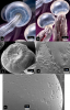
Secretion of resin from glandular heads.
a, b) Highly magnified heads using macrophotography and phase contrast imaging showing distribution of vesicles inside the heads. A ring of secretory cells can be seen on the underside of the heads above the stalk. (Images are courtesy of 11 Zoom Gardens and are included with the acknowledgement of credit to the photographer Nick Cash).
c, d) Scanning electron micrographs of the ring of secretory cells (c) and droplets of resin secreted on the outside of the cuticle (d).
e) Microscopic pores (approx. 0.5 um in diameter) that presumably may allow resin to be secreted through the cuticle. The confirmation of the presence and function of these pores requires additional studies.
 jcannabisresearch.biomedcentral.com
 www.instagram.com
Caption and source link to research below.
Secretion of resin from glandular heads.
a, b) Highly magnified heads using macrophotography and phase contrast imaging showing distribution of vesicles inside the heads. A ring of secretory cells can be seen on the underside of the heads above the stalk. (Images are courtesy of 11 Zoom Gardens and are included with the acknowledgement of credit to the photographer Nick Cash).
c, d) Scanning electron micrographs of the ring of secretory cells (c) and droplets of resin secreted on the outside of the cuticle (d).
e) Microscopic pores (approx. 0.5 um in diameter) that presumably may allow resin to be secreted through the cuticle. The confirmation of the presence and function of these pores requires additional studies.
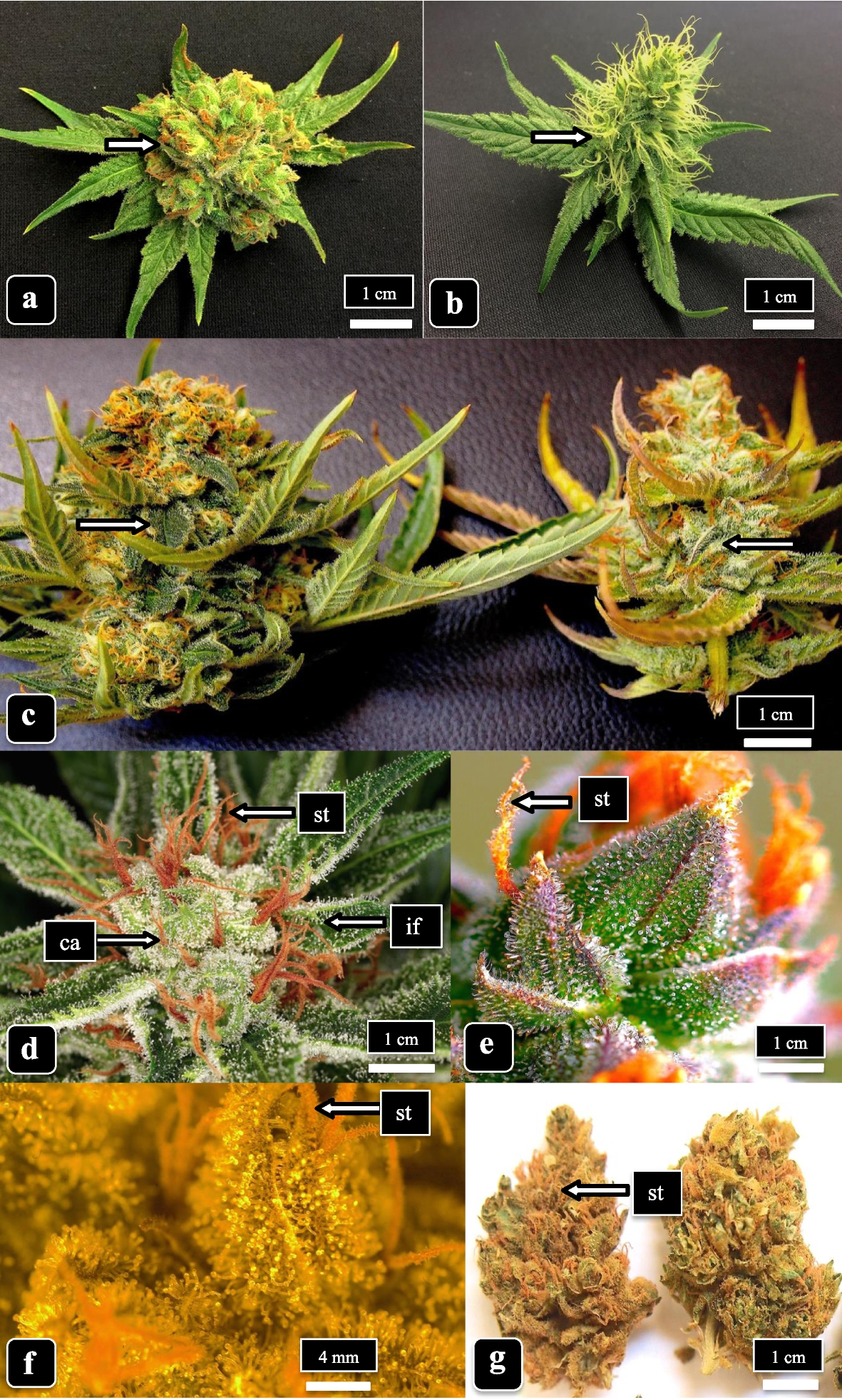
Glandular trichome development, morphology, and maturation are influenced by plant age and genotype in high THC-containing cannabis (Cannabis sativa L.) inflorescences - Journal of Cannabis Research
Background Glandular capitate trichomes which form on bract tissues of female inflorescences of high THC-containing Cannabis sativa L. plants are important sources of terpenes and cannabinoids. The influence of plant age and cannabis genotype on capitate trichome development, morphology, and...
Login • Instagram
Welcome back to Instagram. Sign in to check out what your friends, family & interests have been capturing & sharing around the world.
www.instagram.com
Last edited:
